The human body contains millions of T-cells, each one of them expressing a unique receptor that allows them to mount a highly targeted immune response. This is referred to as the “immune repertoire” and is known to be an important indicator of immune health. Having a wide variety of T-cells increases the probability of having a T-cell receptor that recognizes one of the many pathogens that you may encounter in your life. However, when generating millions of unique T-cell receptors (a process known as VDJ recombination), mistakes will certainly be made.
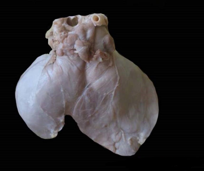
Enter the thymus – a small organ located between our lungs, just behind the sternum. The thymus is responsible for shaping our entire T-cell repertoire by testing out every single one of those millions of unique T-cells. This process is a lot like the story of Goldilocks and the Three Bears, when Goldilocks is trying to find porridge that is the right temperature for her to eat. In this analogy, Goldilocks (the thymus) is trying to eliminate the porridge (T-cell) that is too hot or too cold. Our bodies require T-cells that will react appropriately, and the thymus accomplishes this by getting rid of the T-cells that overreact to normal, healthy proteins in our body (“too hot”) or that don’t react at all (“too cold”).
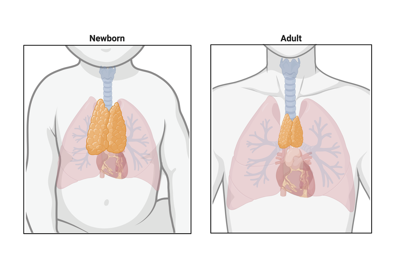
The problem, however, is that the thymus rapidly declines with age. It is the only organ that is larger in childhood than in adulthood. By puberty, the thymus begins to gradually shrink in a process known as “thymic involution”, churning out fewer and fewer T-cells. The result of thymic involution is a significantly smaller, less diverse immune repertoire, causing the elderly to be exceptionally prone to infectious disease, cancer, and autoimmunity. For this reason, scientists have long pondered strategies to regenerate the thymus, with limited success, until now.
Incredibly, for the first time, scientists have successfully built a functioning human thymus capable of supporting the development of functioning human T-cells. Investigators at the Francis Crick Institute and University College London recently published groundbreaking work in Nature Communications providing proof of concept for bioengineering a functioning human thymus. While other bioengineered organs have been generated in the past, the complex nature of the thymus presents a particular challenge.
Going back to our Goldilocks analogy, the thymus gets rid of T-cells that are “too cold” or “too hot” through processes known as positive and negative selection, respectively. First T-cells are positively selected for, meaning that the thymus plucks out all the newly generated T-cell receptors that can properly bind to MHC molecules (MHC molecules are expressed on the surface of cells throughout the body and present small fragments of proteins to T-cells to identify the presence of pathogens). A T-cell receptor that cannot correctly bind to MHC will not be able to mount an immune response when it needs to (“too cold”) and will undergo “death by neglect”. The positively selected T-cells that can bind MHC then continue through the thymus and progress to the next selection step: negative selection, meaning deletion of T-cells that recognize “self”.
This may sound philosophical, but it is a rather important (and straightforward) concept in immunology. Discrimination of “self” versus “non-self” is of utmost importance when thinking about your immune system because we want our T-cells to attack foreign invaders (viruses, bacteria, etc.), but not our healthy tissues and organs. Negative selection accomplishes this feat with the help of a unique (and very cool) protein, AIRE (Autoimmune Regulator). AIRE is specifically expressed by cells within the thymus and allows those cells to generate normal, healthy proteins that are found in every single tissue throughout your body. That means proteins that are only found in your kidney, spleen, or even your eyeball are also found in this little piece of tissue in your chest (cool, right?). These “self” proteins are presented on MHC to the recently positively selected T-cells. Then, any T-cells that mount a strong immune response to these normal, healthy proteins found throughout your body (“too hot”), will be eliminated (i.e. negative selection). Thus, we are left with a T-cell repertoire that will appropriately mount an immune response against foreign pathogens, but will not inappropriately attack healthy tissues throughout our body.
These specialized cells within the thymus that express AIRE (and consequently produce every single protein found in our bodies) are called Thymic Epithelial Cells, i.e. TECs. While epithelial cells are fairly commonplace (lining our skin, intestines, and blood vessels), the ability of TECs to serve these vital functions in the thymus are part of the reason this cutting-edge work is so groundbreaking. The ability to generate sufficient numbers of cells that replicate the highly specialized functions of TECs has been a significant hurdle to bioengineering a functioning thymus. Paola Bonfanti, senior author on the publication, and her team were able to greatly expand human TECs in vitro long-term for the first time. To do this they obtained human thymi donated from patients, thinly sliced them, carefully laid those slices over surgical sponges, and bathed them in culture medium. These tissue fragments could then be enzymatically digested and processed into single cell suspensions, which are capable of growing for an extended period of time with proper nutrients.
Another significant obstacle for Bonfanti and her team was finding an appropriate scaffold (a physical structure or foundation) upon which these expanded TECs may grow. To do this, researcher Asllan Gjinovci developed a novel, micro-surgical technique to remove all non-structural cells from rat thymi while maintaining the 3D anatomy and vasculature. The team was then able to inject the scaffolds with the expanded human TECs, and they found that they were not only able to repopulate the whole-organ scaffolds, but the cells also assembled themselves into the unique epithelial structure of the thymus with functional and supportive cells interwoven. Finally, the team implanted these reconstituted thymi into mice to determine their functionality. Remarkably, the investigators found that these bioengineered thymi were able to support human T-cell development in 77.5% of all implanted thymi.
While the authors note that this system requires further optimization and investigation, it has the potential to propel immunology research forward. It opens the door to greatly enhancing our understanding of mechanisms and factors related to T-cell development, positive and negative selection, and generation and maintenance of tolerance to self. There are also many potential medical applications for this technology. Thymic transplantation could promote tolerance in the context of organ transplantation, reducing the need for life-long immunosuppressive drugs by allowing a mechanism (negative selection by a transplanted thymus) through which the transplant recipient’s immune system can produce T-cells that will not attack the transplanted organ. Thymic transplantation also holds the potential to overcome severely debilitating immunodeficiencies that result from thymic dysfunction. And more broadly, this technology may be used to better understand and combat thymic involution associated with the immune system’s gradual decline with age.
Image information:
“Pleural based thymoma – Cytokeratin AE1/AE3 immunostain – Case 282” by Pulmonary Pathology is licensed under CC BY-SA 2.0
“File:Human thymus.jpg” by Dr. Roshan Nasimudeen is licensed under CC BY-SA 3.0
Figure created by the author with Biorender
Megan Stanley Molina is a doctoral student in the Department of Immunobiology at the University of Arizona. Her research interests include dendritic cells, mechanisms of immune tolerance, and cancer immunotherapy. You can find her on twitter @SciStan or instagram @meganst_nley.


Thank you for this great write up! Your goldilocks analogy fits very well. Shared this on my LinkedIn since I liked this so much 🙂
LikeLike