From 2015 to 2016, Americans watched as a curiously-named virus spread like wildfire throughout South America, leaving a trail of birth defects in its wake. This virus, known as Zika virus, or ZIKV for short, eventually entered the United States, and with it came the fear that these birth defects would start popping up in the US as well. ZIKV is a flavivirus that is initially transmitted via mosquitoes but can be passed between sexual partners as well. The virus quickly garnered a reputation for causing birth defects, most notably microcephaly (the development of undersized heads in newborns). Exactly what causes these adverse outcomes, particularly microcephaly during pregnancy, is still not well understood. However, recent research has uncovered more about the host immune response to this virus during pregnancy. Surprisingly, this work suggests that the immune system’s attempt to eradicate ZIKV might not be as strictly beneficial as we thought.
Antibodies and Antibody-dependent enhancement
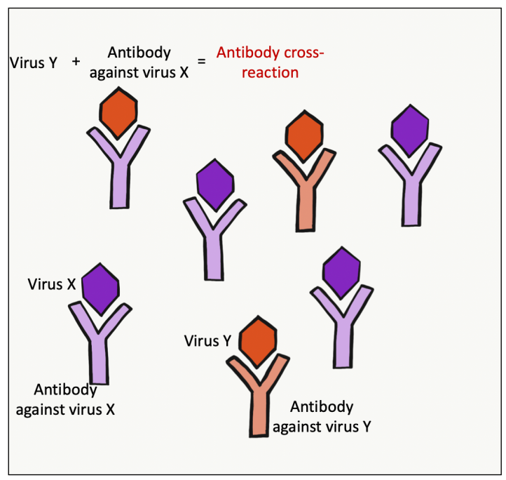
When a virus like ZIKV infects a person, Y-shaped immune proteins known as antibodies are produced by immune cells within the body. These proteins can bind, neutralize, and ultimately help to combat the invading viruses. Antibodies can also remain within the body for extended periods of time, long after the initial infection, to help limit the severity of subsequent infections (this is part of the basis for vaccines). While generally beneficial, there are rare instances in which these antibodies can have a negative effect by cross-reacting.
Antibody cross-reaction occurs when an antibody specific for one virus recognizes another, closely-related virus, and inappropriately binds to it (Figure 1). Because this bound antibody is not specific for the closely-related virus, it cannot fully neutralize and combat the pathogen. Instead, the bound antibody allows for the closely-related virus to enter into cells it wouldn’t otherwise have had access to, thus helping to increase virus infection. Like most immune system misregulation, it’s a case of good intentions gone wrong.
This enhancement of a viral infection by cross-reactive antibodies is known as antibody-dependent enhancement (ADE) and most notably occurs between members of the mosquito-borne Flaviviridae family like ZIKV and dengue virus (DENV). In areas where mosquito-borne viral infections are common, the development of ADE becomes a prominent health concern. To address this, Rathore et al. set out to explore if and to what extent ADE could play a role in ZIKV pathogenesis, particularly in the development of microcephaly.
The role of ADE in microcephaly and fetal development
To model a scenario in which ADE could occur, the authors first infected mice with DENV and then infected the same mice with ZIKV three weeks later. Compared their non-DENV infected counterparts, mice that received both ZIKV and DENV infections produced much smaller pups with growth-restricted heads. In a similar setup, mice were injected with antibodies for DENV and then infected with ZIKV. The fetal growth restrictions and birth defects were even more severe in these animals, suggesting that maternal DENV antibodies (not the DENV itself) were playing a key role in the severity of ZIKV-associated birth defects. To confirm these results, the authors measured different markers of proper neuronal development in the mice. They observed less of these markers in mice that received either DENV or DENV antibody treatment prior to ZIKV infection compared to non-treated mice. This suggests that the presence of DENV antibodies during subsequent ZIKV infection negatively influences proper brain development by restricting neuronal development in the fetuses.
Despite the clear birth defects, it remained unclear whether any ZIKV was directly infecting the pups and causing these adverse outcomes or whether the maternal immune response was entirely to blame. Upon further examination, the authors discovered that the virus was able to infiltrate both the placenta and the fetus and that it was actively replicating within these tissues. Additionally, mice that had DENV antibodies prior to ZIKV infection had increased viral load within their fetuses. Placental cells within these prior challenged mice also showed an increased presence of ZIKV proteins. Taken together, these results suggest that the presence of DENV antibodies actually increased direct ZIKV infection of fetuses. This supports the notion that ADE has a role in potentially worsening the associated adverse outcomes and birth defects associated with ZIKV during pregnancy.
Neonatal Fc receptor function and misuse
While it was clear that prior DENV infection, specifically the antibody response, was enhancing the pathology of subsequent ZIKV infection, questions of how this was happening still lingered. To piece this together, the authors turned to an antibody receptor that has been previously implicated in cases of ADE called the neonatal Fc receptor (FcRN). This receptor, which is a protein that sits on the surface of cells and grabs onto other proteins, like antibodies, is present on a wide range of tissues. Most notably, the FcRN is found on the surface of various immune cells. The classic function of FcRN is to bind the stem of Y-shaped antibodies (known as the Fc region) and allow for the entry of antibody-coated and neutralized virus into immune cells like macrophages. Once inside the immune cell, the neutralized virus is degraded, thereby preventing its spread.
Given the important role of FcRN in mediating the degradation of viral particles by immune cells, you would think that this protein would globally dampen viral infection. In practice, though, this is not always the case, and when ADE is at play, the rules of the game change. Specifically, in the case of DENV-enhanced ZIKV infection, previously-produced antibodies specific for DENV bind to the invading ZIKV but cannot completely neutralize it. This antibody-ZIKV complex gets taken up by immune cells, but since ZIKV is not neutralized, the virus is able to continue replicating within the immune cell. This, in turn, promotes its accumulation and spread within the body. Moreover, when the infected person is an expecting mother, the impact of FcRN-mediated ADE could be more severe (Figure 2).
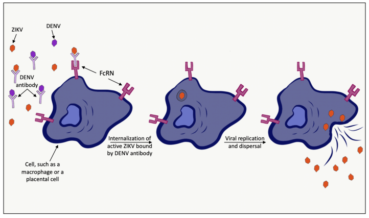
In addition to being present on immune cells, FcRN is also present on the cells of the placenta, where it allows for the transfer of maternal antibodies to the developing fetus. While this function is normally invaluable to early immune system development in the fetus, it could potentially have disastrous effects when a virus like ZIKV infects the mother. More specifically, it’s possible that maternal antibodies could be binding to ZIKV and allowing for its entry into the placenta and fetus via the FcRN. If this were the case, then FcRN would not only be mediating ZIKV immune evasion and expansion, but it would also allow this high viral load to be passed on to the developing fetus.
To elucidate if FcRN is involved in the DENV-enhanced ZIKV infection of fetuses, the authors utilized mice that completely lacked FcRN (also known as FcRN knockout mice) and conducted similar experiments as those described above. By deleting FcRN from these mice, scientists were able to determine a necessary role for FcRN in ADE. As in previous experiments, knockout mice were infected first with DENV, then three weeks later with ZIKV. Interestingly, the FcRN knockout mice had similar levels of ZIKV within the fetus whether or not they were infected with DENV previously. This suggests that FcRN is potentially mediating the ADE of ZIKV infection in the fetus. However more work is needed to fully understand this complex system.
Some final thoughts
The underlying implications of these findings are critical. They not only help to shed light on the mechanism of ZIKV-associated microcephaly but also suggest that individuals in areas where both DENV and ZIKV are endemic are at greater risks of developing ZIKV-associated birth defects. As such, these findings drive home the need for better treatment options for those doubly-affected and at risk individuals.
Cover image: Zika by Nick Youngson CC BY-SA 3.0 Alpha Stock Images
