Thousands of skilled assassins are currently moving through your body. Natural Killer cells, or NK cells, are constantly on the watch, checking up on cells to make sure they are healthy and have not gone rogue. If a cell is healthy, the NK cell passes it by. But if it is infected or cancerous, the NK cell kills it, swiftly and precisely.
How much ammunition does a NK cell carry around? Exactly how much does it use on a cell it is trying to kill? And, most importantly, how much ammunition does it really need to kill one cell? Recently, scientists from the Baylor College of Medicine have examined precisely these questions, discovering new details of NK cell killing mechanisms.
Degranulation – Taking a Closer Look
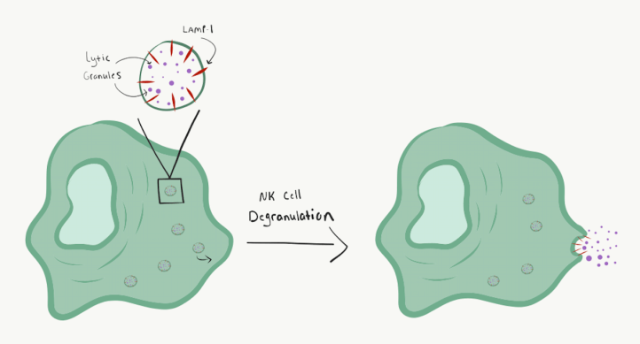
It was previously known that NK cells kill by releasing small packages of enzymes, called lytic granules, onto a target cell. As the name implies, these lytic granules kill other cells by poking holes in the cell membrane of the target cell, causing the cell to lyse or burst. Prior to expulsion from the NK cell, the lytic granules group together where the NK cell is touching the target cell, and then the membranes of the granules fuse with the cell membrane, releasing their contents (Figure 1). This process is called degranulation.
More recently, scientists found that while NK cells have around 200 lytic granules per cell, they only release less than 20 granules onto target cells. This wasn’t a huge shock, as NK cells are known “serial killers,” with the ability to kill one target cell and immediately move on to kill another. As such, it makes sense that they would not dispel all of their granules at once, allowing them to sequentially attack multiple cells without waiting to replenish their granule stocks. More surprising, however, was that NK cells only need to release 2-4 lytic granules to kill a target cell.
How were scientists able to determine this? They needed a way to visualize both the death of the target cells and the degranulation of the NK cells.
Cell death can be visualized using many different methods, but these scientists chose an indicator dye called calcein-AM that would be present in live cells but would quickly exit cells that die. As such, the color of the cells serves as an indicator of their viability, with live cells maintaining the dye and dead cells being colorless. Compared with other methods for looking at cell death, it works very quickly. A sensitive indicator was important for these experiments, as they were trying to determine a minimum number of lytic granules that could cause cell death.
To visualize degranulation events, the scientists made use of a membrane protein called LAMP-1 (lysosomal-associated membrane protein-1). LAMP-1 is part of the membrane of lytic granules, spanning the membrane with a short tail in the cytoplasm and the majority of the protein inside the lytic granule. The membrane of the lytic granule is much like the membrane of the cell, though much smaller in scale, and serves to contain the enzymes so they do not digest the NK cell itself. When a lytic granule releases its contents, its membrane fuses with the cell membrane, and the inside of the lytic granule ends up on the outside of the cell. (Figure 1) When this happens, LAMP-1 goes from the very acidic inside of the granule to the less acidic cell membrane and surrounding environment.
Taking advantage of this change in pH, scientists fused LAMP-1 with a pH-sensitive fluorochrome, called pHlourin. Fluorochromes are small molecules that absorb light of one wavelength and emit light of a different wavelength. When in the acidic environment of the lytic granule, pHlourin’s fluorescence is masked, but in neutral pH conditions of the exterior cell membrane, it fluoresces under green light. By using cells that expressed the fusion of LAMP-1 to pHlourin, the scientists could visualize degranulation events by the appearance of this fluorescence.
Using these two methods of visualization and time-lapse imaging with high-resolution microscopy, the scientists were able to look at live NK cells as they attacked target cells, counting single degranulation events until the point of cell death.
Why bother? – The importance of basic science
What good is understanding the exact mechanics and numbers behind NK cell degranulation? These experiments are an example of basic science, which tries to understand how biology works. Other types of scientific investigation are translational research, which tries to apply basic science concepts to medicine, and clinical research, which is even more directly applicable to human patients. Why focus on basic science when there are other types of research that seem more directly applicable to human health?
Biology is incredibly complex. Even one system within it, such as NK cells, is like a huge intricate machine with all kinds of interlocking and interacting gears. When we want to manipulate that system, for example, with a potential new drug, we are trying to change how one of those gears interacts with the rest to alter the outcome. But if we don’t understand the basic biology of NK cells, it would be like working in a dark room where we can’t see the machine.
Basic science experiments shine spotlights on this machine. Some of them might illuminate a large portion, and some a portion that’s very small—even the size of just a few lytic granules—but every spotlight helps us understand how the parts of the machine work together. The better we understand the whole, the better we will be able to understand the effects of manipulation such as therapeutic drugs and avoid unintended consequences on human health.
After all, if every cell in our body is as complex as a machine, imagine the human body as a factory, full of many different machines that all have to work together for a common purpose. Altering one machine in a unpredictable way can result in side effects that were not anticipated. The more we know, the more minutely and carefully we can make adjustments to the individual machines of our cells to improve the overall function of the factory that is our bodies.
NK Cells in Cancer Immunotherapy
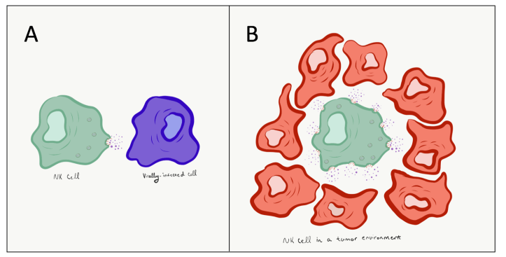
How can a better understanding of NK cells apply to health-related scenarios? Since one of the functions of NK cells is to kill tumor cells, many people have looked at using NK cells in cancer immunotherapy. Several clinical trials are currently in progress using NK cells in different ways to treat multiple types of cancer. So far, treatment with NK cells shows a good safety profile with no observed toxicity, but their ability to kill tumor cells has been mild.
In the discussion of their paper, these scientists proposed a way their basic science experiments could potentially lead to better outcomes for NK cells as immunotherapeutic agents when treating tumors. When an NK cell degranulates, it brings its lytic granules into a focused area, exactly where it is touching the target cell. This is a very efficient method when killing one specific cell, but NK cells in a tumor environment are surrounded by potential target cells (Figure 2). If NK cells could be directed to degranulate from multiple locations across its membrane’s surface, they could potentially be more efficient at killing tumor cells. How can we manipulate NK cells to have this unfocused degranulation? Another experiment for another day!
This article was based upon the following study: Gwalani, et al. Single Degranulations in NK Cells Can Mediate Target Cell Killing. J Immunol. 2018 March 28; ji1701500 (2018)
The cover image was generated by the National Institute of Allergy and Infectious Diseases.
